Structure of Gallbladder
The gallbladder is a pear shaped sac, which lies in a depression present on the under surface of the liver. It is about 7.5 – 12.5cm in length; with a capacity of about 30-50ml. Anatomically the gallbladder can be divided into three parts; a fundus, body and a neck.
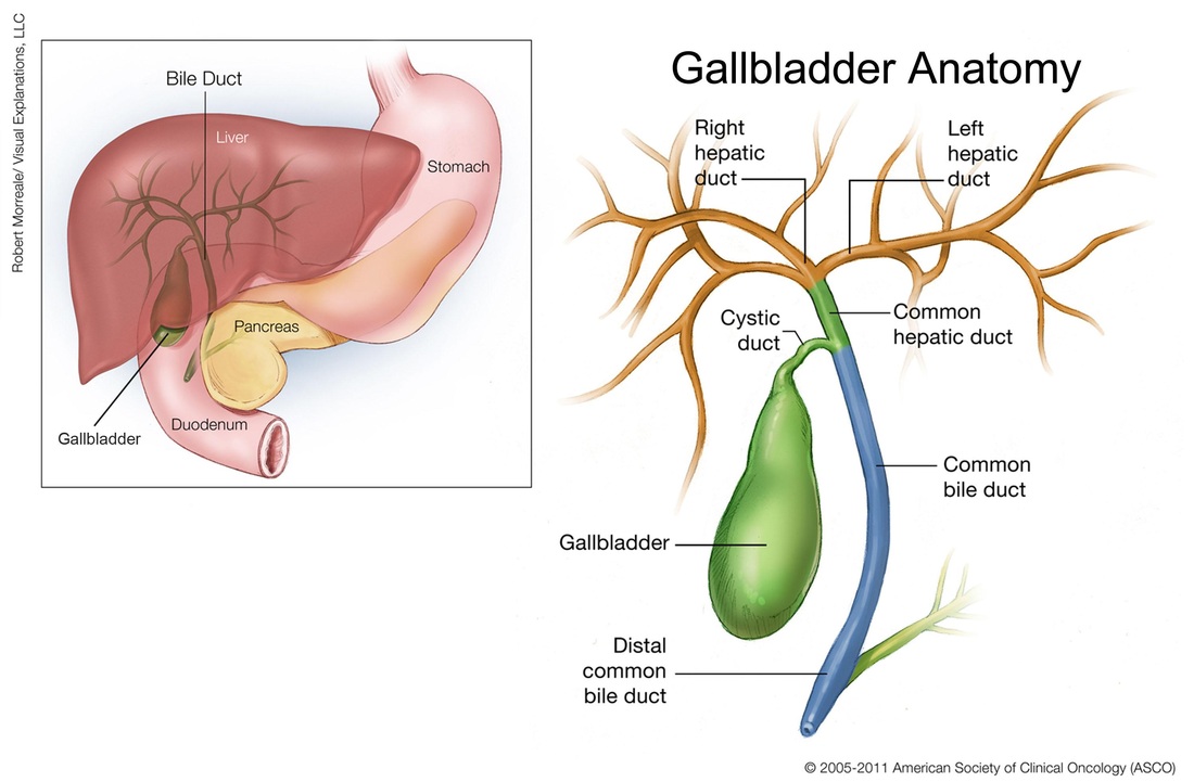
Fig.1 Anatomy of the gallbladder. n.d. [online] Available at:<http://www.cancer.net/multimedia/medical-illustrations-gallery?mitid=232>[Accessed 20 May 2013]
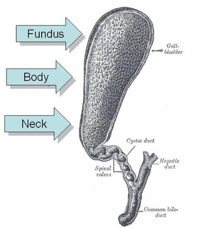
Fig.2 Parts of a gallbladder. n.d. [online] Available at:<https://www.healthtap.com/#topics/gallbladder-diseases-bile-duct-diseases> [Accessed 20 May 2013]
Relations of the Gallbladder
Anterior to (in front of) the gallbladder is the liver and abdominal wall.
Posterior to (behind) the gallbladder lies the transverse colon (part of large intestine) and 1st and 2nd part of duodenum (first part or division of small intestine).
Posterior to (behind) the gallbladder lies the transverse colon (part of large intestine) and 1st and 2nd part of duodenum (first part or division of small intestine).
Anatomy of the Ducts
The cystic duct is a 2.5cm long, S-shaped narrow tube which is continuous with the neck of the gallbladder. It has a valve called the spiral valve of Heister which keeps the (lumen) duct open and joins the common hepatic duct which is less than 2.5cm in length, (common hepatic duct is formed from right and left hepatic ducts coming from the liver) to form the common bile duct. The common bile duct is approximately 7.5cm in length. It joins the pancreatic duct (coming from the pancreas) and dilates to form an ampulla called the ampulla of Vater, which finally opens into the wall of the duodenum, (part of the small intestine) by means of a papilla called the major duodenal papilla. The ampulla is surrounded by a circular smooth muscle called the sphincter of Oddi.
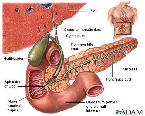
Fig.3 Anatomy of the biliary tree. n.d. [online] Available at:<http://www.umm.edu/patiented/articles/what_gallstones_gallbladder_disease_000010_1.htm> [Accessed 20 May 2013]
Vessels and Nerves
Arterial supply of the gallbladder is by the cystic artery, which is a branch of hepatic artery and is usually given off behind the common hepatic duct.
Venous drainage is through the cystic vein which empties into the portal vein.
Lymph from the gallbladder goes to cystic lymph nodes, present near the neck of the gallbladder, which drain into the hepatic nodes along the course of hepatic artery and finally into the celiac nodes.
Nerve supply to the gallbladder is by the celiac plexus. Contraction of the gall bladder is under the influence of the hormone cholecystokinin (CCK), which is produced from the mucous membrane of the duodenum.
