Investigations
Investigations carried out to confirm the diagnosis are:
Blood Tests
Blood Tests
- It shows an increased white blood cell WBC or total leukocyte count TLC (raised in infection and inflammation)
- Increased levels of bilirubin, Alkaline phosphatase and serum aminotransferases (liver enzymes)
Ultrasonography
It is a non-invasive, painless technique; considered the best test for detecting gallstones. It uses high frequency sound waves to visualise the tissues of the body. It demonstrates the biliary stones, size and wall thickness and inflammation of the gallbladder. It can also detect stones in the bile duct. There are no complications of ultrasound; however it is of less value in patients who are obese and those who have a lot of air in their intestines.
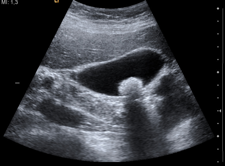
Fig.7 Ultrasound showing single large gallstone. n.d. [online] Available at:< http://commons.wikimedia.org/wiki/File:Ultrasound_image_of_gallbladder_stone_Gallstone_091937515.jpg> [Accessed 20 May 2013]
|
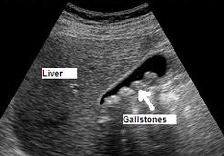
Fig.8 Multiple gallstones in a gallbladder. n.d. [online] Available at: < http://www.ourwebdoctor.com/gallstones.htm> [Accessed 12 May 2013]
|
X- Rays
It shows radiopaque gallstones in 10% of the patients and calcification of the gallbladder.
Intravenous Cholangiography
A solution called biligram is injected intravenously, which is rapidly secreted by the liver into the biliary tree. Subsequent radiography clearly defines the duct and gallbladder, delineating the presence of disease.
Computerised Tomography CT
Computerized tomography is of less value for detecting gallstones in the gallbladder; but demonstrates dilated bile ducts, which indicates the presence of stones. It is mainly used for detecting complications of gallstones.
MRI and MRCP
Magnetic resonance imaging MRI, takes picture of internal organs, with a powerful magnet, which produces images from inside the body. Magnetic resonance cholangiopancreatography MRCP is a relatively new technique, which gives a clear and detailed outline of the biliary tree and the presence of gallstones in the ducts, using special software combined with MRI technology. It has no complications but the downside is that patients with metallic implants or devices are advised not to take the test.
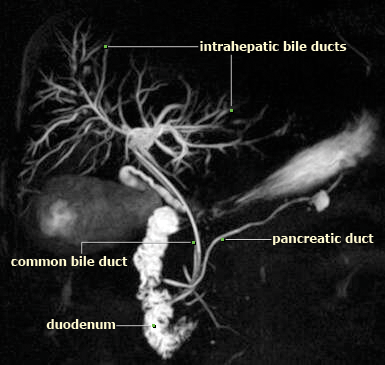
Fig.9 MRCP showing the biliary tract. n.d. [online] Available at: <http://imaging-mri.blogspot.co.uk/2012/11/mrcp-imaging-anatomy.html> [Accessed 20 May 2013]
ERCP
Endoscopic retrograde cholangiopancreatography ERCP is the gold standard technique for detecting gallstones in the bile ducts. It is diagnostic as well as a therapeutic technique. The procedure involves mildly sedating the patient so that the endoscope can be inserted through the mouth without irritating the patient. The tube goes through the stomach to reach the intestine. At this point a water soluble, contrast dye is injected via a tube through the endoscope. The dye travels up (retrogradely) within the biliary tree and X-ray images are taken of the biliary and the pancreatic ducts. This procedure thus allows the physician to see two sets of images i.e. the endoscopic view of the intestine as well as detailed X-rays of the biliary tree and the pancreatic duct.
For therapeutic action and complications of the procedure refer to ERCP discussed under treatment of gallstones in bile ducts.
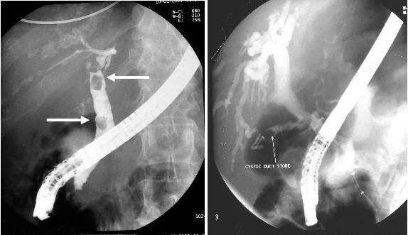
Fig.10 ERCP images, left: stones in CBD and right: stone in cystic duct. n.d [online] Available at:<http://www.ceessentials.net/article41.html> [Accessed 12 May 2013]
EUS
Endoscopic ultrasonography EUS is a new, low-risk procedure using high-frequency sound waves during endoscopy. EUS allows the physician to study the gallbladder, pancreas, and bile ducts and to accurately detect gallstones in the common bile duct.
